Biomedicum Imaging Unit Faculty of Medicine |
|
|
Contact information _______________________
5th floor office, room B501a 1st floor office, room B117a Biomedicum Helsinki
BIU web site: |
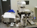
Zeiss LSM 780 confocal microscope
Technical Information
Microscope Motorized Zeiss Axio Observer.Z1 inverted microscope Detectors
Objectives
Laser lines
Fluorescence filters for VIS-mode
Transmitted light
Software Zeiss ZEN 2010 software. Physiology, FCS for GaAsP, ROI HDR, and Image Correlation Spectroscopy analysis packages for ZEN 2010. Accessories for live cell and multiwell imaging
Instrument Use
General instructions If you would like to start using the LSM 780, please go to our Fees and user policy page for instructions for new users. Important safety issue: avoid looking at the laser light - the lasers are much more powerful than the ones in laser pointers and may be harmful for your eyes. After your session, please check if users after you on the same day will need the same lasers as you. If yes, leave them on (argon in standby). If not, turn the lasers off. When you start Internet Explorer in the computer, it will automatically open the scheduler (our online reservation system) page that shows all the same-day reservations for LSM 780. As all users should mark the laser lines they are planning to use (in Description), you can easily check if you should leave the lasers on, or turn them off. If you are the last user of the day and you leave the lasers on, you will be charged for the whole time the lasers are on. Turn on the lights when you clean up, otherwise you may not notice oil spills and other problems. Cleaning up - instructions what to do after you are done with imaging. In case of an accident - instructions what to do if you spill oil or break glass. The last user of the day should turn off the computer; first log off, then choose log off again, select the option shut down. You can open ZEN files in your own computer (provided that it is PC, not Mac) using Zeiss ZEN lite. This freeware can be downloaded from the Zeiss web site. All registered LSM 780users are automatically added to the LSM 780users mailing list. Please read those e-mails to know what's going on! Links
Molecular Expressions. Excellent microscopy reference for beginners and experts by M.W. Davidson and the Florida State University. Olympus has excellent web pages on confocal microscopy, full of useful information on fluorophores, spectral bleed-through, resolution and contrast, objectives etc. Highly recommended. Based on Molecular Expressions. Nikon has also an outstanding website that offers a weath of information on confocal microscopy, as well as on wide-field microscopy. Learn about sample preparation, aberrations, and other important aspects of confocal imaging. Based on Molecular Expressions. University of Buffalo is maintaining confocal listserv. You can subscribe the confocal list (you'll get all the e-mails sent to the list and you can send your own ones) or you can search the list archives without subscription. The list is one of the best ways of learning about advanced confocal microscopy.
Page updated 21.10.2016 |
||||||||||||||||||||||||||||||||||||||||||||