Biomedicum Imaging Unit Faculty of Medicine |
|
|
Contact information _______________________
5th floor office, room B501a 1st floor office, room B117a Biomedicum Helsinki
BIU web site: |
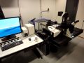
Leica TCS CARS SP8 confocal microscope
Technical Information Microscope Motorized DMI 6000 inverted microscope Detectors
Objectives and their properties
Single photon laser lines
Multi photon laser
Accessories for live cell and multiwell imaging
Instrument Use
General instructions If you would like to start using SP8, please go to our Fees and user policy page for instructions for new users. Important safety issue: avoid looking at the laser light - SP8's lasers are much more powerful than the ones in laser pointers and may be harmful for your eyes. Especially the picoEmerald emits invisible, high powered IR light that can cause permanent damage to your eye-sight. Turn on the lights when you clean up, otherwise you may not notice oil spills and other problems. Cleaning up - instructions what to do after you are done with imaging. As the environmental chamber and the microscope take a long time to cool, the first reservation made for a particular day will determine the temperature used on that day. In other words, the temperature cannot be changed during the day. When you reserve the SP8, note the temperature you'll be using into the "Description" field. You can abbreviate it as "RT" for room temperature and "37" for +37 Centigrade. Check the previous reservations for the day when you're making your own and copy the temperature note so users after you don't have to guess. In case of an accident - instructions what to do if you spill oil or break glass. All registered SP8 users are automatically added to the SP8 users mailing list. Please read those e-mails to know what's going on! Please contact in advance if you need temperature adjustment for your experiments.
Links
Molecular Expressions. Excellent microscopy reference for beginners and experts by M.W. Davidson and the Florida State University. Olympus has excellent web pages on confocal microscopy, full of useful information on fluorophores, spectral bleed-through, resolution and contrast, objectives etc. Highly recommended. Based on Molecular Expressions. Nikon has also an outstanding website that offers a weath of information on confocal microscopy, as well as on wide-field microscopy. Learn about sample preparation, aberrations, and other important aspects of confocal imaging. Based on Molecular Expressions. University of Buffalo is maintaining confocal listserv. You can subscribe the confocal list (you'll get all the e-mails sent to the list and you can send your own ones) or you can search the list archives without subscription. The list is one of the best ways of learning about advanced confocal microscopy.
Page updated 21.10.2016 |
|||||||||||||||||||||||||||||||||||||||||||||||||||||||||||||||||||||||||||||||||||||||||